Characterise the cell death pathway or protection mechanisms of your compound
Cell death is a necessary physiological process, used to eliminate damaged and excess cells to preserve homeostasis and maintain healthy tissues. By assessing cell death morphology and the cascade of biological events leading to cell death, it is possible to explore stress and post-trauma responses, and compounds that can prevent or reduce organ dysfunction in these contexts.
Our MitoXpert® and MitoPathway® platforms characterise cell death mechanisms through a large panel of cellular models.
Identify compounds with efficacy on tumour cells and characterize cell death pathways.
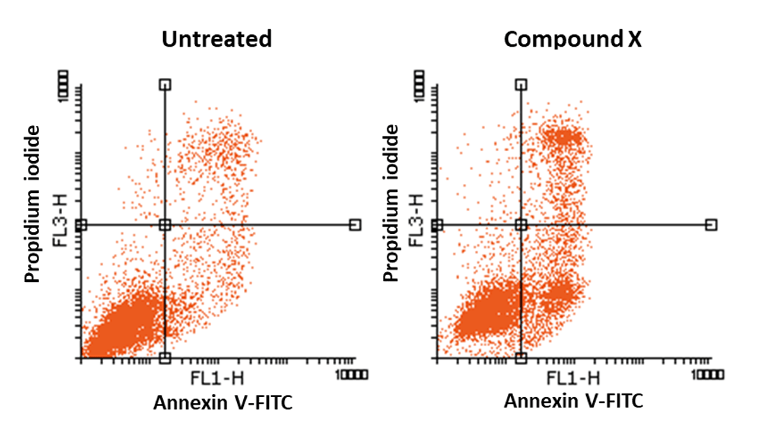
Analysis of apoptosis induction in BxPC3 pancreatic cancer cells by measuring plasma membrane permeabilization (Propidium iodide labeling) and phosphatidylserine externalization (Annexin V-FITC labeling) by flow cytometry. This method allows distinction between necrosis (PI +/AV -) from primary (PI -/AV +) and secondary apoptosis (PI +/AV +).
Whereas cell death is a physiological process, acute accidental cell death – in case of ischaemia, for example – or accumulation of dead cells within a tissue in response to a range of stressors inducing inflammation or fibrosis can lead to serious organ dysfunction or even loss of complete areas.
Cancer cells are particularly resistant to cell death, and recent therapeutic strategies aim to activate mitochondrial cell death pathways in tumour cells, for example by targeting the Bcl-2 family members to trigger apoptosis, or mPTP to induce necrosis, or by modulating mitophagy. To improve the efficacy of targeted therapies and prepare the way for potential drug combinations, it is of particular interest to characterise drug-induced cell death mechanisms in cancer cells.
Using MitoXpert® and MitoPathway®, you can characterise cell death mechanisms to:
- Demonstrate a protective effect of compounds in pathological toxicity contexts, including metabolic stress exposure, ischaemia-reperfusion injury due to oxygen and/or glucose deprivation,
- Understand the mechanisms through which compounds provide cell protection in disease models, and reveal potential mitochondrial target(s),
- Identify compounds targeting tumour cells and characterise the cell death pathway – apoptosis, necrosis, autophagy-dependent cell death – induced by anti-tumour compounds.
Evidence mitochondrial pro-apoptotic features of anti-tumour compounds.
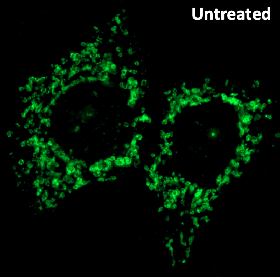
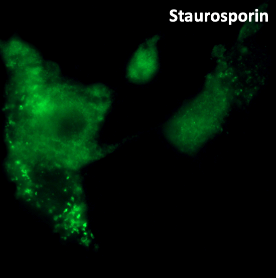
Induction of cytochrome c release (apoptosis marker) in BxPC3 tumor cell line by treatment with the pro-apoptotic agent staurosporin. Fluorescent microscopy is performed after labeling with anti-cytochrome c antibody.